It's not just about what you see, but how you see it
The human eye, an intricate marvel of biology, allows us to perceive the world in all its vibrant hues and intricate details. However, beneath the surface of this remarkable organ lies a complex network of structures that work in harmony to provide us with clear vision. Among these structures is the retinal pigment epithelium (RPE), a single layer of specialized cells that plays a crucial role in maintaining healthy vision.
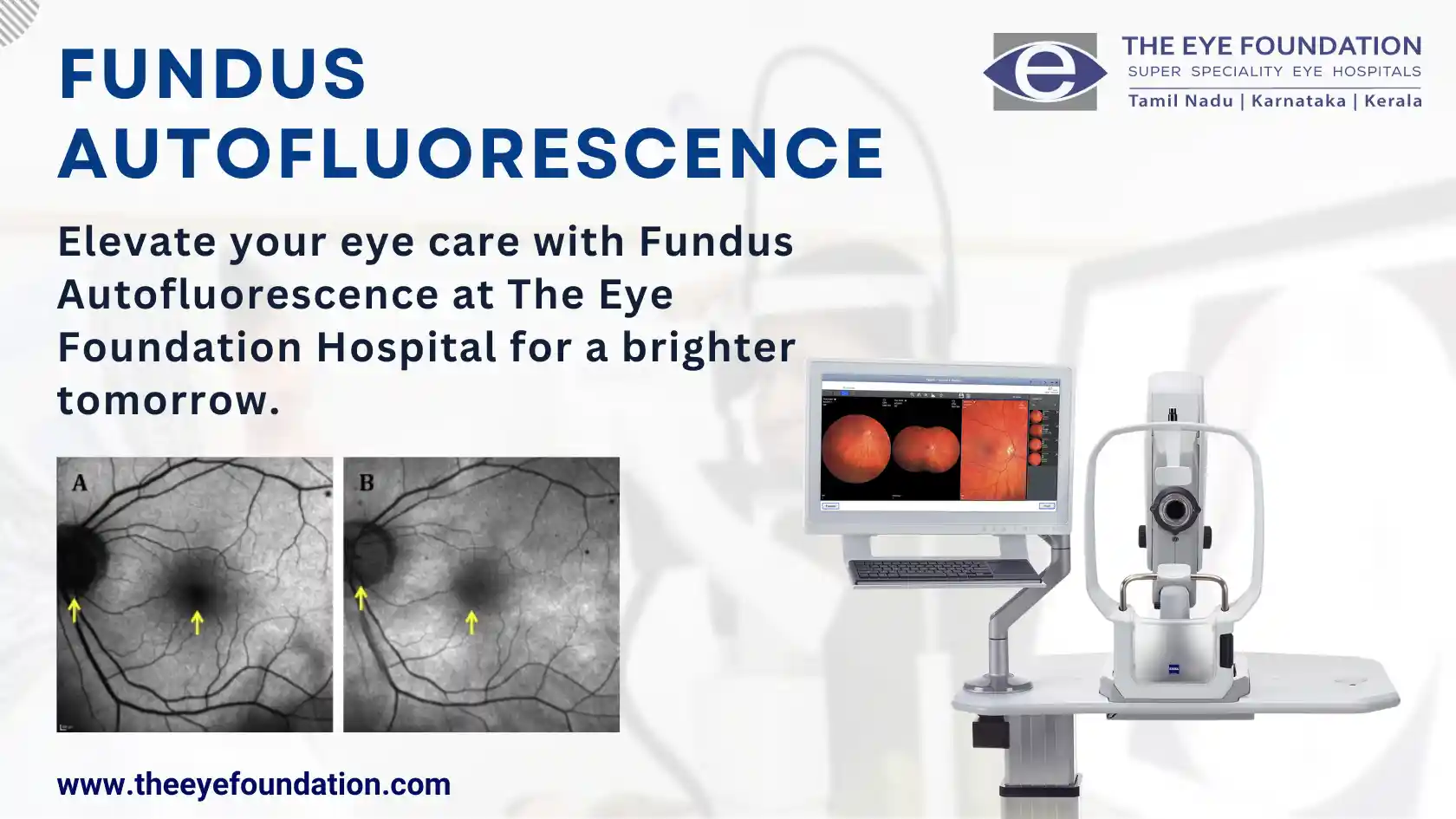
Fundus autofluorescence (FAF) is a non-invasive imaging technique that unveils the health and function of the RPE by illuminating naturally occurring fluorescent molecules within its cells. These molecules, known as fluorophores, emit light when exposed to a specific wavelength of blue light, providing a valuable window into the RPE's health and the presence of underlying retinal disorders.
Unraveling the Mysteries of RPE Health
FAF imaging offers a unique perspective on RPE health by revealing patterns of autofluorescence that can vary depending on the individual's retinal condition. Increased autofluorescence, for instance, may indicate the accumulation of lipofuscin, a by-product of cellular metabolism. While lipofuscin is a normal part of aging, excessive accumulation can signal retinal stress or damage.
On the other hand, decreased autofluorescence may suggest a loss of RPE cells, a hallmark of various retinal diseases. By discerning these subtle variations in autofluorescence patterns, FAF imaging provides valuable insights into the health and integrity of the RPE.
A Diagnostic Tool for Retinal Disorders
FAF imaging has emerged as a powerful diagnostic tool for a wide spectrum of retinal disorders, including:
- Age-related macular degeneration (AMD): FAF can detect early signs of AMD, such as drusen deposits and geographic atrophy, facilitating early intervention and potentially preventing vision loss.
- Diabetic retinopathy: FAF can identify subtle changes in fluorescence associated with diabetic retinopathy, aiding in monitoring disease progression and guiding treatment decisions.
- Retinal pigment epithelium (RPE) disorders: FAF can reveal abnormalities in autofluorescence distribution, helping to diagnose RPE-specific conditions such as multifocal choroiditis and atrophic RPE lesions.
The Eye Foundation: Your Gateway to Clear Vision
At The Eye Foundation, we understand the importance of early detection and intervention in retinal disorders. Our team of experienced vitreo-retinal specialists utilizes cutting-edge FAF imaging technology to provide comprehensive diagnostic evaluations and personalized treatment plans for patients with a wide range of retinal conditions.
Book an Appointment Today
If you are concerned about your eye health or have a family history of retinal disorders, we encourage you to schedule an appointment with our vitreo-retinal specialists at The Eye Foundation. Our dedicated team is committed to helping you achieve clear vision and preserve your precious gift of sight.


