 Tamil Nadu
 Karnataka
 Kerala

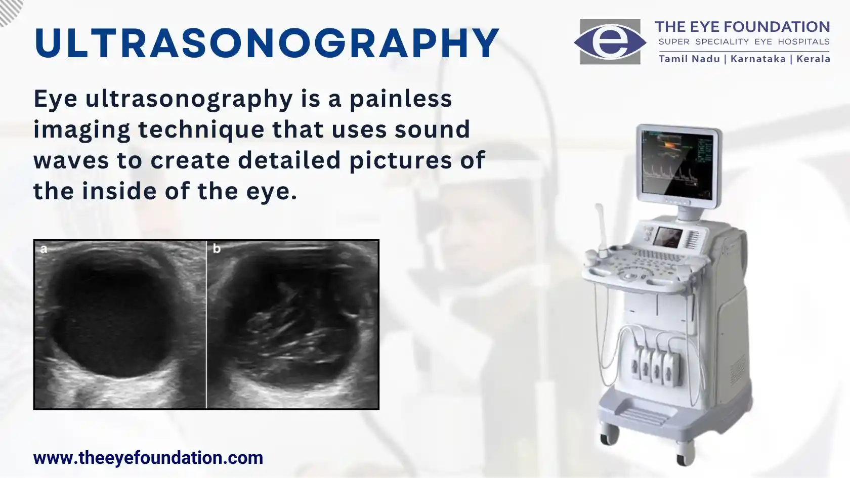
Unveiling the Invisible with Sound Waves
Ultrasonography, also known as an eye ultrasound, is a painless and non-invasive diagnostic tool that utilizes high-frequency sound waves to create detailed images of the internal structures of the eye. This versatile technique plays a crucial role in evaluating a wide range of eye conditions, providing valuable insights for diagnosis, treatment planning, and monitoring disease progression.
Principles of Ultrasonography
The principle underlying ultrasonography is based on the reflection of sound waves as they encounter different densities of tissue within the eye. The echoes produced by these reflections are captured and converted into detailed images, revealing the intricate anatomy of the eye's interior.
Types of Ultrasonography
There are two primary types of ultrasonography commonly used in ophthalmology:
- A-scan ultrasound: This technique measures the axial length of the eye, a crucial parameter for determining the appropriate intraocular lens (IOL) power in cataract surgery.
- B-scan ultrasound: This technique produces cross-sectional images of the eye, providing detailed visualization of the vitreous cavity, retina, optic nerve, and other posterior segment structures.
Applications of Ultrasonography
Ultrasonography offers a versatile diagnostic tool for a wide range of eye conditions, including:
- Evaluating retinal detachment: Ultrasonography can effectively detect retinal detachments, even in the presence of opaque cataracts or vitreous hemorrhage.
- Assessing vitreous abnormalities: Ultrasonography is valuable for visualizing vitreous opacities, foreign bodies, and intraocular tumors.
- Measuring ocular tumors: Ultrasonography can accurately measure the size and location of ocular tumors, aiding in treatment planning.
- Guiding vitreoretinal procedures: Ultrasonography can provide real-time guidance during vitreoretinal surgeries, ensuring precise surgical maneuvers.
Ultrasonography at The Eye Foundation
At The Eye Foundation, our experienced ophthalmologists are well-versed in performing and interpreting ultrasonography. We utilize state-of-the-art ultrasound equipment to obtain high-resolution images, providing comprehensive diagnostic information for our patients.
Book an Appointment
If you have concerns about your eye health, schedule a consultation with our vitreoretinal specialists at The Eye Foundation. We will conduct a thorough examination, including ultrasonography if necessary, to accurately diagnose your condition and recommend the most appropriate treatment plan.